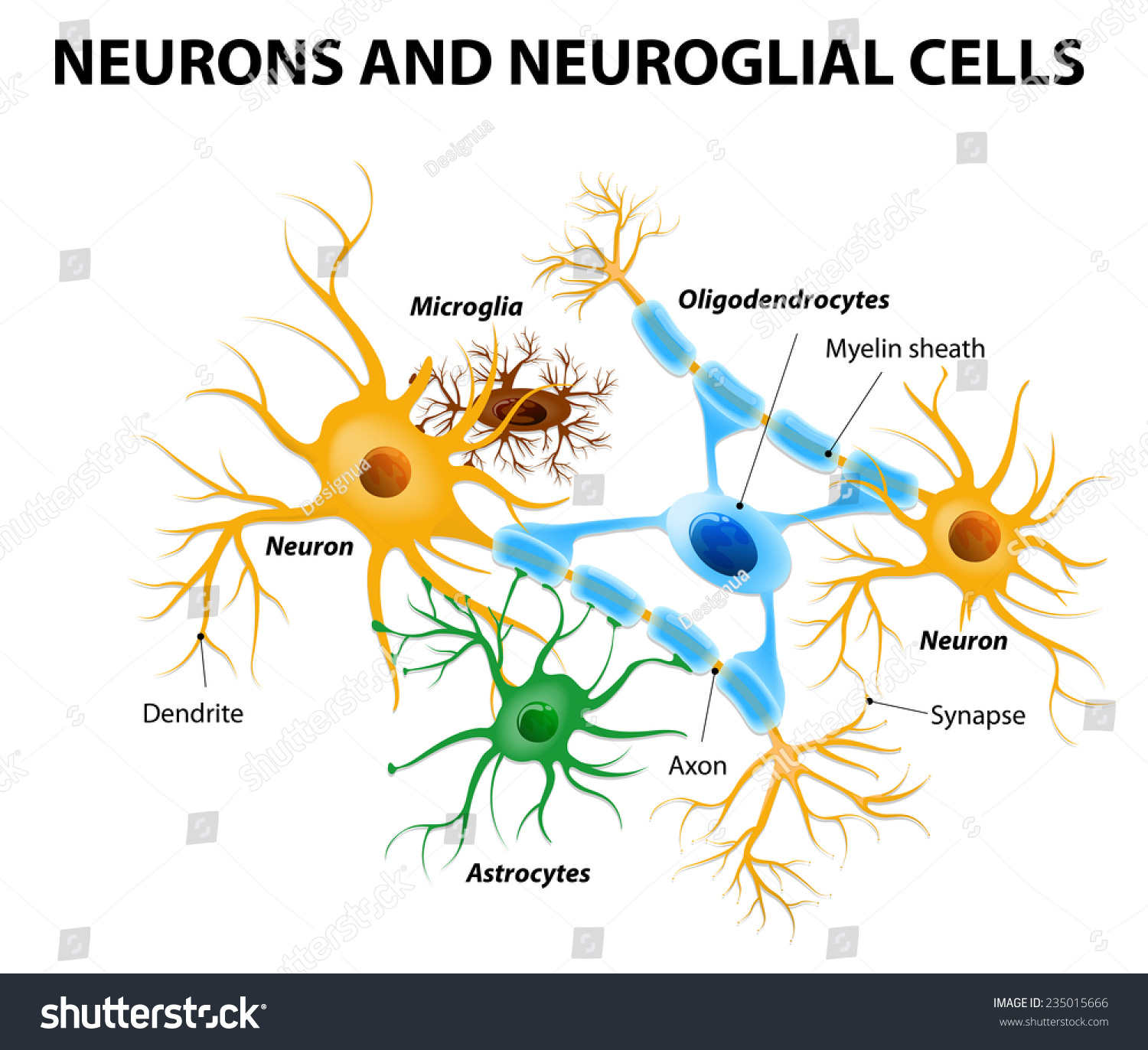
Neurons Neuroglial Cells Glial Cells Nonneuronal Stock Vector 235015666
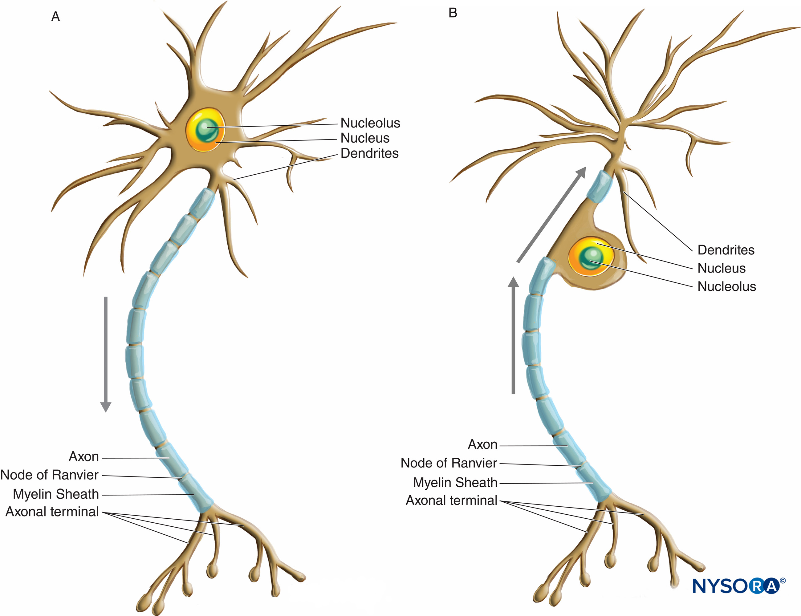
Like other cells, each neuron has a cell body (or soma) that contains a nucleus, smooth and rough endoplasmic reticulum, Golgi apparatus, mitochondria, and other cellular components. Neurons also contain unique structures, illustrated in Figure 35.3 for receiving and sending the electrical signals that make neuronal communication possible.

Color the Neuron and Neuroglial Cells Answer Key CameronkruwMccann
The neuron is one of two basic types of cells in the nervous system, the other type being the glial cell. Figure 11.3.1 11.3. 1: Interneurons of Adult Visual Cortex. Neurons, also called nerve cells, are electrically excitable cells that are the main functional units of the nervous system. Their function is to transmit nerve impulses.
Color the Neuron and Neuroglia
Overlaid cells in t-SNE plots with marker genes for SGC and Schwann cells show the relative levels of expression as a color. neuroglial cells and number and volume of nerve cells in the spinal.

Nervous Tissue Mediates Perception and Response Anatomy and Physiology I
Color the Neuron and Neuroglia Students can practice what they have learned about neurons with this simple coloring activity. The page shows features of the neuron, such as the axons and dendrites. They will also color the supporting cells of the matrix.
Color The Neuron And Neuroglial Cells Worksheet Master
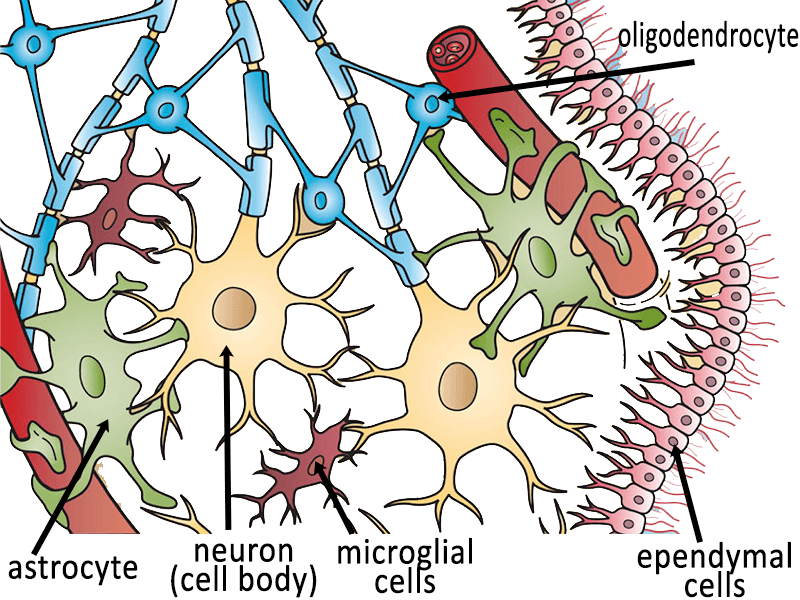
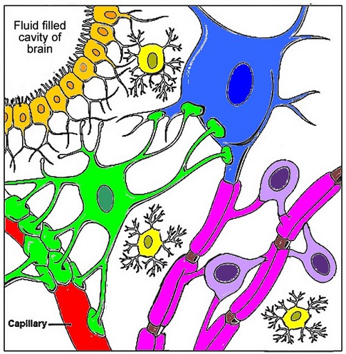
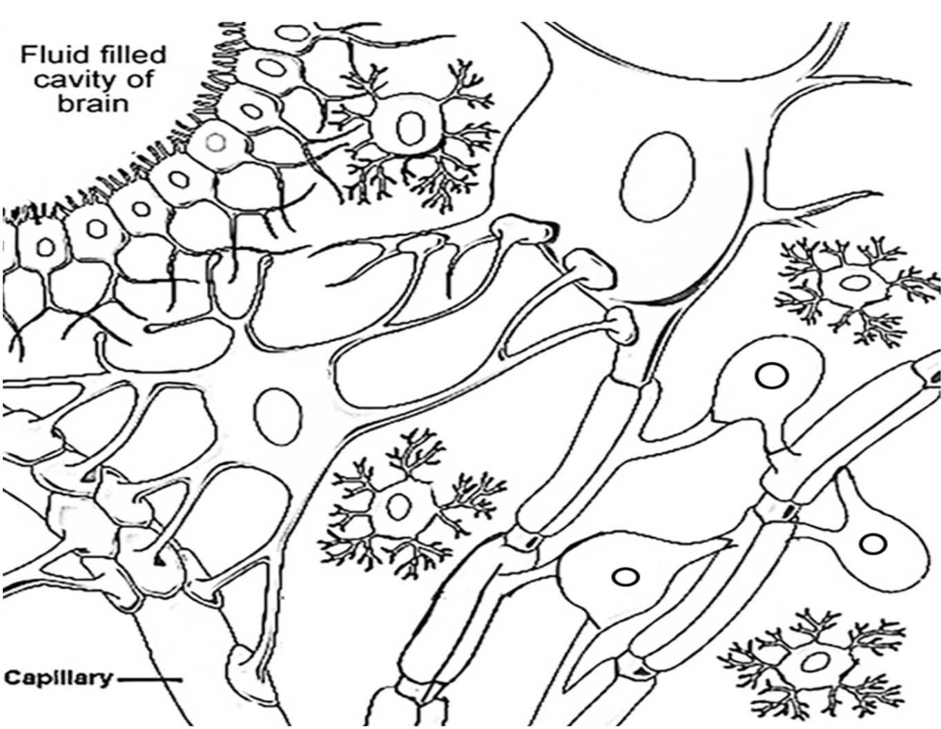
For each of the cells above, color the nucleus a darker shade of purple, green, blue, orange Myelin sheaths (pink) Capillary (red) Microglial cells (yellow) Nodes or Ranvier and the Axon (brown) What is the function of: 1) Oligodendrocytes _______ make the myelin sheaths ______________________________
Histology of the Peripheral Nerves and Light Microscopy NYSORA
astrocyte: a neuroglial cell, in the shape of a star, in the brain. Neurogila or glial cells, are non-neuronal cells that maintain homeostasis, form myelin, and provide support and protection for neurons in the central (CNS) and peripheral nervous systems (PNS). It was long believed that neuroglia did not play any role in neuro-transmission.

Medical Terms & Сlinical Сases Neuron
Neuroglia cells are different from nerve cells in that they do not participate directly in synaptic interactions Students can also label a nerve cell and color neuroglia cells using paper handouts. I have also had students model neurons with clay and chalk markers.

Color The Neuron And Neuroglial Cells
1.1. The Birth of the Concept of Homoeostatic Neuroglia. The complexity of the human brain is remarkable: a population of more than 200 billion (i.e. 2 × 10 11) neural cells (neurones and neuroglia) is packed within a limited volume (average human brain occupies 1200-1400 cm 3).These neural cells form complex networks, connected through 15-20 trillions of chemical and electrical synapses.
Neurons and Glial Cells · Biology
Glial Cells. Nervous tissue is composed of two types of cells, neurons and glial cells. Neurons are the primary type of cell that most anyone associates with the nervous system. They are responsible for the computation and communication that the nervous system provides. They are electrically active and release chemical signals to target cells.

Color The Neuron And Neuroglial Cells
Like the heart, lungs, and stomach, the nervous system is made up of specialized cells. These include nerve cells (or neurons) and glial cells (or glia ). Neurons are the basic functional units of the nervous system, and they generate electrical signals called action potentials, which allow them to quickly transmit information over long distances.

Types of neurons. Structure sensory, motor neuron, astrocyte
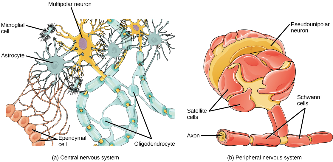
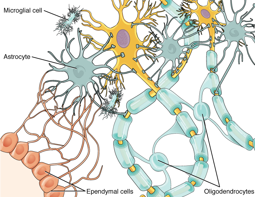
Figure 35.3.1 35.3. 1: Glial cells: Glial cells support neurons and maintain their environment. Glial cells of the (a) central nervous system include oligodendrocytes, astrocytes, ependymal cells, and microglial cells. Oligodendrocytes form the myelin sheath around axons. Astrocytes provide nutrients to neurons, maintain their extracellular.
19+ Art Labeling Activity Neuroglial Cells Of The Cns MarilyamShae
List and describe the functions of the structural components of a neuron Compare the functions of different types of glial cells Explain the stages of an action potential and how action potentials are propagated Explain the similarities and differences between chemical and electrical synapses

Color The Neuron And Neuroglial Cells
Neuroglia, also called glia or glial cells, are non-neuronal cells of the nervous system. They compose a rich support system that is essential to the operation of nervous tissue and the nervous system. Unlike neurons, glial cells do not have axons, dendrites, or conduct nerve impulses.Neuroglia are typically smaller than neurons and are about three times more numerous in the nervous system.

Anatomy of the Nervous System Microbiology
Today, we recognize three broad groups of glial cells: (i) true glial cells or macroglia, such as astrocytes and oligodendrocytes, of ectodermal origin, the stem cell of which is the spongioblast; (ii) microglia, of mesodermal origin; and (iii) ependymal cells, also of ectodermal origin and sharing the same stem cell as true glia.

Neuron and Neuroglial Cells 8131534 Vector Art at Vecteezy
neuroglia, any of several types of cell that function primarily to support neurons.The term neuroglia means "nerve glue." In 1907 Italian biologist Emilio Lugaro suggested that neuroglial cells exchange substances with the extracellular fluid and in this way exert control on the neuronal environment.It has since been shown that glucose, amino acids, and ions—all of which influence.
Color the Neuron and Neuroglial Cells Biology LibreTexts
Color the Neuron and Neuroglial Cells Oligodendrocytes (purple) Astrocyte (green) Ependymal Cells (orange) Body of Neuron (blue) Myelin sheaths (pink) Capillary (red) Microglial cells (yellow) Nodes or Ranvier and the Axon (brown) What is the function of: 1) Oligodendrocytes ____________________________________________
